Spirochete Research Labs

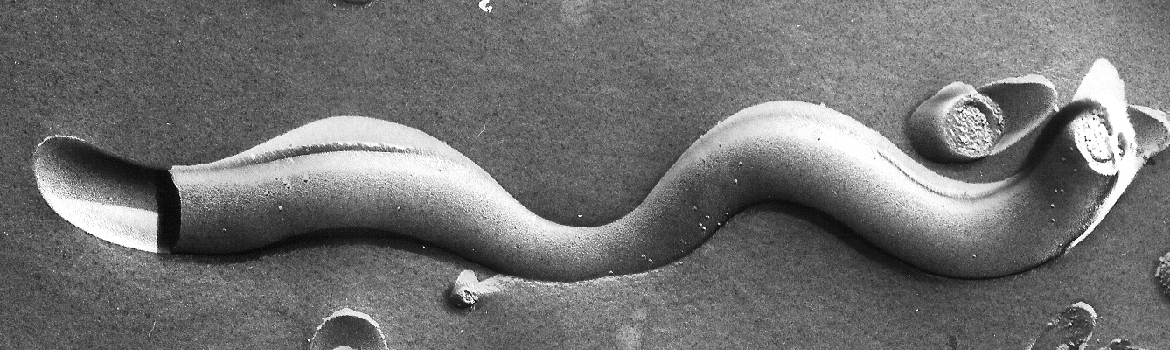
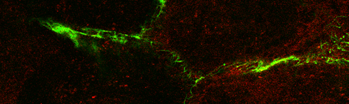
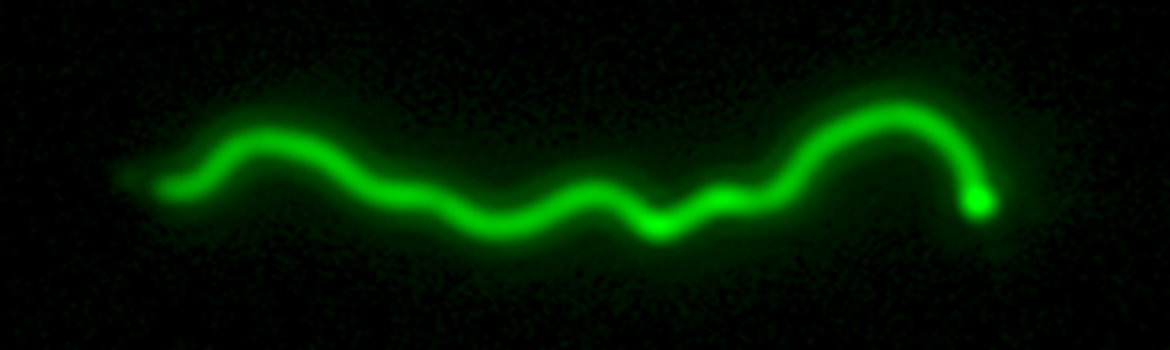
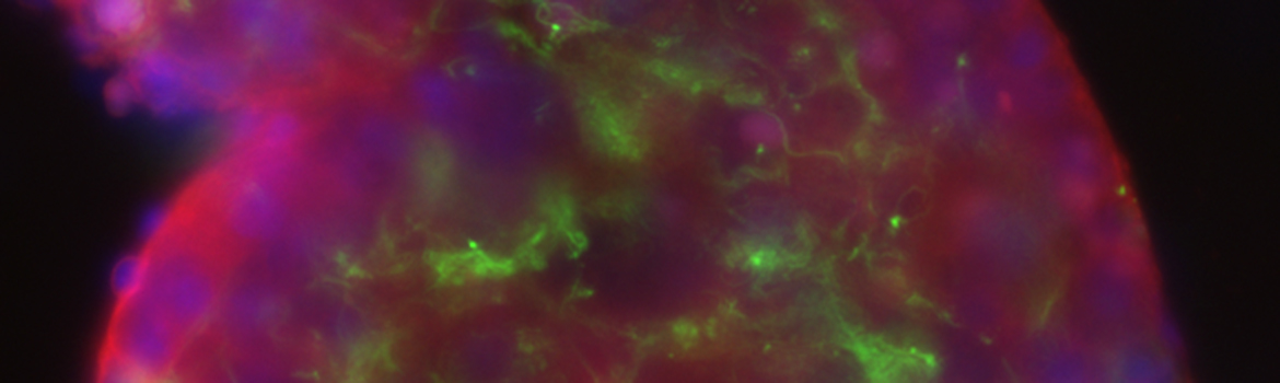
Spirochetes are an ancient and extremely successful eubacterial phylum characterized by distinctive helical or planar wave-form morphology and flagellar filaments confined to the periplasmic space. Spirochetes from the genera Leptospira, Treponema, and Borrelia are highly invasive pathogens that, as the agents of leptospirosis, syphilis (T. pallidum), Lyme disease (B. burgdorferi), and relapsing fever (B. hermsii, B. recurrentis, and others), pose public health problems of global dimensions. T. denticola and numerous other treponemal species, most of which remain uncultivated, are major components of the polymicrobial biofilms that cause periodontal disease. In recent years, the availability of genomic sequences for a number of spirochetes, improved methodologies for genetically manipulating these fastidious bacteria, and novel light and microscopic technologies have provided powerful new tools for studying the ultrastructures, genetics and parasitic strategies of these unusual pathogens.

Lyme Disease/Borrelia burgdorferi

Radolf Laboratory
Photo of tick specimens
The Lyme disease spirochete B. burgdorferi is maintained in nature via an enzootic cycle in which it is transmitted by the nymphal stage of its vector, the deer tick Ixodes scapularis, to a mammalian host (typically the white-footed mouse Peromyscus leucopus) and then is acquired when a naïve larval tick feeds on an infected mouse. Infection of humans with B. burgdorferi is accidental and is not required for persistence of the spirochete in nature. In order to transit between its arthropod and mammalian hosts, spirochetes must decipher complex environmental cues delivered at the feeding site and, in response, undergo dramatic changes in their transcriptomes and proteomes. The principal objective of Lyme disease research conducted in the Radolf Laboratory is to understand these processes. The alternative sigma factor RpoS is a unifying genetic feature of this project. Signals delivered by the blood meal induce the expression of RpoS which, as the promoter-reading subunit of RNA polymerase, induces far-reaching changes in the bacterium’s transcriptome: (1) upregulation of approximately 100 B. burgdorferi genes that we believe are required for transmission of spirochetes from tick to mouse and/or the establishment of infection once within the mouse and (2) downregulation of approximately 30 tick-phase genes that spirochetes no longer need once they are inoculated into mice.

Critical to this work has been our development of green fluorescent protein (GFP) reporters that enable us to track live spirochetes in ticks and mice. Our live-imaging studies have fundamentally changed our understanding of the transmission process. In order to reach the mouse, spirochetes disseminate through the midgut into the salivary glands in order to access the salivary stream which they “ride” into the vertebrate host. We have found that dissemination of spirochetes in ticks is actually biphasic. In the first phase, which we have termed “adherence-mediated migration, spirochetes replicate in close association with differentiating midgut epithelial cells, “working” their way as aggregates or networks to the base of the epithelium. In the second phase, they transition into typically motile spirochetes, complete the penetration through the midgut, and then move on to the salivary glands en route to the mouse. Most recently, we have found that spirochetes lacking RpoS are deficient in this process and we are developing various strategies to identify the RpoS-dependent genes involved.

Salazar Laboratory
Since Bb lacks orthologs of known exotoxins or the specialized secretory machinery required for the delivery of noxious molecules into host cells, it is widely accepted that the distinctive inflammatory clinical signs and symptoms associated with the disease result from the human host’s innate and co-evolving adaptive immune responses to the bacterium. Monocytes and macrophages are considered to be two critical cellular elements of the innate immune response to Bb. Innate immune recognition of the spirochete by these cells was previously thought to result primarily from the interactions of the bacterium’s abundant outer membrane-associated lipoproteins with CD14 and Toll-like receptors (TLR) 1/2. However, experimental evidence generated in the Radolf-Salazar laboratories has demonstrated that phagocytosed Bb induces inflammatory signals that differ both quantitatively and qualitatively from those generated by lipoproteins at the cell surface of monocytes/macrophages and dendritic cells. In our 2009 PLoS Pathogens publication (Salazar et al), we demonstrated that internalized spirochetes also induce transcription of interferon-β (IFN-β) and several type I interferon-stimulated genes (ISGs). The finding that spirochetal lipoproteins were unable to induce type I IFNs confirmed that in the case of Bb these responses occur independently of TLR2.

Photo of researchers looking into microscopesRecent evidence from the Salazar laboratory, published in the Proceeding of the National Academy of Sciences (Cervantes et al.), indicates that the recognition of borrelial ligands by the monocyte involves a cooperative interaction between TLR2 and TLR8 in the generation of pro- and anti-inflammatory cytokine responses, whereas endosomal TLR8 is solely responsible for IRF-7 mediated induction of IFN-β. The role of TLR8 in generating type I IFNs and the cooperative nature of TLR2 and TLR8 signaling at the phagosome, which we have designated “phagosomal signaling,” had not been previously described or examined in a human model. Our combined observations have enabled us to formulate a new model for innate immune recognition of LD spirochetes. In this model, binding of unopsonized Bb to the monocyte/macrophage cell surface, through a yet to be characterized phagocytic receptor, sets the stage for the broader immune signaling events that follow and which mechanistically can only be integrated following internalization of the bacterium and formation of the phagolysosome. As maturation of the phagosome proceeds, this unique vacuolar structure appears to be capable of sensing diverse spirochetal PAMPs, becoming an increasingly more efficient platform for signal generation which sets the stage for enhanced production of TLR2 and TLR8-mediated NF-ĸB mediated pro- and anti-inflammatory cytokines and TLR8-dependent induction of IFN-β.

Syphilis/Treponema pallidum

The syphilis spirochete T. pallidum harbors many resemblances to B. burgdorferi but actually employs a markedly different parasitic strategy. Whereas B. burgdorferi is an enzootic pathogen, T. pallidum is an obligate pathogen of humans which cannot be cultivated in artificial medium. The modes of transmission of the two bacteria differ markedly as well: T. pallidum is transmitted from person-to-person by sexual activity, whereas B. burgdorferi is transmitted by an arthropod vector. Once within the host, T. pallidum begins to replicate locally, eventually causing a genital ulcer, called a chancre, the clinical hallmark of the primary stage of the disease. As the chancre develops, treponemes begin to make their way towards draining lymph nodes and blood vessels in order to spread systemically. Once within the blood, T. pallidum is extremely adept at invading virtually every organ system in the body, including the central nervous system, and establishing persistent infection that can cause serious, even life threatening, complications months to years later. We have designated T. pallidum “the stealth pathogen” because of its remarkable ability to evade host immune defenses.

Radolf Laboratory
Efforts in the Radolf Laboratory to explain T. pallidum’s stealth pathogenicity have focused on the bacterium’s unusual molecular architecture. Over the years, we have generated abundant evidence that the T. pallidum outer membrane differs markedly in structure and composition from its gram-negative counterparts (e.g., Escherichia coi). Not only does it lack lipopolysaccharide, the highly inflammatory glycolipid in the outer membranes of gram negative bacteria, it also contains a much lower density of integral membrane proteins that present few surface antigenic targets to the host immune system. Situated below the outer bilayer, where they are inaccessible to antibodies in intact organisms, are the bacterium’s major immunogens, many of which are periplasmic proteins tethered by N-terminal lipids to the cytoplasmic membrane. This work ushered in what we have termed “the quest” for T. pallidum outer membrane proteins, a project that has been ongoing for more than 20 years. Why a quest? Because identifying rare outer membrane proteins is so difficult and requires extraordinary commitment. Fortunately, we now have much more powerful tools to fulfill the quest, among which is the complete genomic sequence of T. pallidum. Genome mining, however, isn’t as easy as it sounds because, with one exception, there are no proteins in the T. pallidum genome with sequence relatedness to well characterized outer membrane proteins of gram-negatives. This work is complicated further by the fragility of the treponeme’s outer membrane. Our genereal strategy is to use bioinformatics algorithms to identify outer membrane protein candidates that then must be cloned, expressed, purified, structurally characterized, and localized in live treponemes. Why is this quest important? Two reasons. First, outer membrane proteins provide channels through which bacteria obtain nutrients. Second, we believe that these surface-exposed proteins, few as they are, are likely vaccine candidates. Given the explosive increase in syphilis cases in the United States and the world during the past decade, a vaccine would be a major weapon in our battle against this centuries old affliction of humans.

In order to fulfill its genetic destiny as a stealth pathogen, T. pallidum must acquire nutrients in every milieu within its obligate human host in which it finds itself, while fending off the host’s attempts to undermine its homeostasis. Recognition of this metabolic war between pathogen and host led us to explore other facets of T. pallidum virulence. One has been transition metal acquisition. Transition metals, such as iron, manganese, and zinc, are essential for life but are present in mammalian body fluids at exceedingly low concentrations. Bacterial pathogens, T. pallidum being no exception, employ highly developed strategems to obtain these nutrients. Our work along these lines has centered about characterizing two ABC transporters within the cytoplasmic membrane (Tro and Znu) that work cooperatively to meet the bacterium’s metal requirements. Lastly, our immune system uses toxic compounds called reactive oxygen species, to kill bacteria. T. pallidum has extremely robust enzymatic mechanisms for detoxifying reactive oxygen species. Understanding how these enzymes work and are regulated in response to host defenses is relevant to all bacterial diseases, not just syphilis.

Salazar Laboratory
Photo of Dr. Salazar discussing his research with Dr. Cruz
The clinical manifestations of venereal syphilis reflect the propensity of Tp to disseminate systemically and to induce chronic inflammation in diverse tissues and organ systems. The appearance of the syphilitic chancre during primary syphilis typically only emerges 2 to 4 weeks after the initial contact with the spirochete. By this time, organisms have disseminated to other organs and a tissue, including the skin, setting the stage for what is classically known as secondary syphilis (SS). This stage of the disease, which is the focus of our translational syphilis research, is characterized by the most overt systemic clinical features including a variety of dermal manifestations as well as systemic signs and symptoms, typically only appearing within 4 to 10 weeks of the initial infection. Paradoxically, despite the robust nature of the adaptive cellular and humoral immune responses typical of SS, which includes the emergence of high titers of opsonic antibodies, it takes weeks and in some cases months for host defenses to gain control of the invading pathogen; ultimately giving rise to an asymptomatic stage known as latent syphilis. How the bacterium is able to evade human host defenses for extended periods of time, while at the same time evoking vigorous cellular and humoral adaptive immune responses during the secondary stage of the disease, is the principal scientific objective of our syphilis translational research program.

A careful analysis of Tp unique ultrastructural features provides several potential explanations for the paradoxical nature of secondary syphilis. Unlike the outer membrane of gram-negative bacteria, that of Tp lacks the potent proinflammatory glycolipid lipopolysaccharide (LPS). In addition, freeze fracture microscopy studies have shown that the spirochete contains very few outer membrane integral membrane proteins (OMPs). Although Tp does contain an abundance of highly antigenic hydrophilic polypeptides, these molecules are tethered by covalently bound N-terminal lipids to the bacterium’s periplasmic inner membrane leaflet. This unusual topology enables intact spirochetes to avoid sensing by innate immune receptors (i.e. Toll-like receptors) present on tissue macrophages and dendritic cells (DCs), while protecting the surface of the organism from the high titers of anti-treponemal antibodies that are generated during SS. Inefficient antibody binding to the sparse spirochetal OMP opsonic targets is thought to allow a large proportion of spirochetes to shun antibody binding and opsonophagocytosis; a mechanism which we have previously demonstrated is an essential requirement for T. pallidum driven innate immune cell activation. Fortunately for the host, a progressively more robust adaptive immune response, including enhanced uptake and degradation of opsonized spirochetes by tissue based macrophages, eventually leads to control of bacterial replication and lesion resolution. Our ongoing translational studies are designed to elucidate the duality of immune evasion and immune recognition of the syphilis spirochete as it occurs in SS, and which builds upon recent advances from the Radolf laboratory in understanding the spirochete’s unique ultrastructure and composition and the host antibody responses it elicits in its natural human host.

To provide an explanation for the paradoxical nature of SS we have proposed a new syphilis immune-pathogenesis model, where antibodies to T. pallidum’s rare outer membrane proteins are only capable of binding a sub-population of spirochetes, thus allowing immune escape of substantial numbers of organisms and at the same time recognition and bacterial clearance of a different subset of spirochetes from blood and tissues. Over time, the host’s expanding repertoire of antibodies against these rare OMPs broadens and intensifies, leading to more efficient opsonophagocytosis by tissue based macrophages, which together with an expanded cell mediated immune response leads to bacterial clearance and lesion resolution. To study key aspects of this model mechanistically, we have developed collaboration with Dr. Adriana Cruz at CIDEIM, a highly prestigious research institute located in Cali, Colombia.